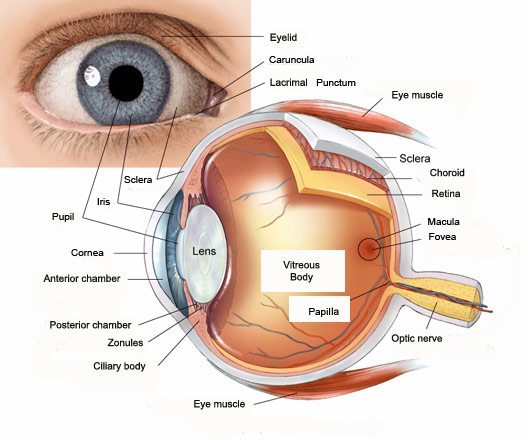
Structure of the eye
The white of your eye is called the sclera covered by the thin conjunctiva. This is a protective layer which covers the entire eyeball except the cornea. The cornea is transparent and it allows light to enter the eye.
The coloured part of your eye is called the iris. The pupil appears as the dark central part of the eye. The lens in the eye can change shape and helps to accommodate for different distances.
The retina is the light-sensitive layer of the eyeball, found on its back wall. The optic nerve carries the electrical impulses created in the retina to the brain.
Most of the eye is filled with a substance like jelly called the ‘vitreous humour’. The front of the eye is filled with a clear fluid called ‘aqueous humour’, which is more watery.
The movement of each eye is controlled by six muscles that pull the eye in various directions. Levator muscle opens the upper eyelid. Orbicularis Oculi muscle closes the eyelid when we blink.

Eyelashes help to stop debris and direct sunlight from entering the eyes by producing a tear film.
The tears come from the lacrimal gland placed just above, and to the outer side, of each eye. The tears then drain down small channels (canaliculi) on the inner side of the eye into a tear ‘sac’. From here they flow down a channel called the tear duct into the nose.
How does the visual system work?
When you look at an object you see it because light reflects off the object and converges (to focus or come together) on the layer of the eye called the retina.
Firstly light passes through the transparent cornea. Most bending of light occurs here. Light then travels through the pupil and hits the lens. The lens also bends light, increasing the amount focused on the highly specialized cells of the retina.
The retina is made up of millions of light sensitive nerve cells called photoreceptors. These cells help to turn the light into electrical signals which are sent to the brain via the Optic nerve to be interpreted in the visual cortex. The visual cortex is a specialised part of the brain which processes visual information. Located at the back of the head, it interprets the electrical signals to get information about the object’s colour, shape and depth. Other parts of the brain put this information together to create the whole picture.

Specialist Ophthalmologist
Bahrain Specialist Hospital
Email: shreyas.palav@bsh.com.bh

